What is Gynecomastia?
What is gynecomastia and how is it distinguished from chest fat?
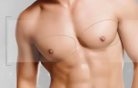
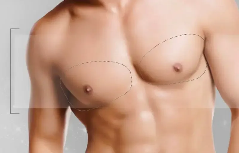
Gynecomastia is the enlargement of the male breast glandular tissue; it presents as a firm or rubbery tissue felt behind the nipple. Fullness caused only by fat accumulation is called pseudogynecomastia; in gyms people often refer to gland enlargement as “gyno.” In many men both conditions coexist and enlargement may be unilateral or bilateral at different degrees. Which tissue is predominant is determined by examination and, if necessary, ultrasound—not by mirror inspection.
Gynecomastia surgery reduces glandular and fat tissue and corrects chest contour; however it does not remove the underlying cause. The chest muscle structure, rib cage shape and natural differences between the two sides are not completely altered by surgery. Information on fat outside the chest is available on the liposuction page; breast reduction performed for large, heavy breasts in women is a different operation (breast reduction).
What causes gynecomastia and which doctor should be consulted?
Gynecomastia is often related to changes in the balance of estrogen and testosterone in the body. Hormonal changes in the newborn period, during adolescence and in older age, certain medications, anabolic steroids and hormone-containing products, excess weight and some hormonal, liver or kidney diseases can contribute to enlargement; in many people no clear cause is found. Investigating the cause prevents missing a treatable medical condition and reduces the risk of recurrence after surgery. Assessment is often a joint effort of two specialties: endocrinology if a hormonal cause is suspected, and plastic, reconstructive and aesthetic surgery if surgical reduction of the tissue is required. Do not stop a prescribed medication on your own because you think it causes enlargement; discuss this with the prescribing physician.
Which symptoms require medical evaluation first?
Medical evaluation is performed before aesthetic planning if symptoms suggest a condition other than gynecomastia. Gynecomastia is typically a central, potentially tender tissue located directly behind the nipple; the following findings warrant additional investigation:
- A firm, irregular, unilateral mass located away from the nipple
- Nipple retraction, bloody or spontaneous discharge from the nipple
- Skin tethering, ulceration or redness on the chest
- Palpable lymph node in the axilla
- Swelling or palpable firmness in the testes
- Rapidly enlarging change over a short period or persistent pain
How do grade and method determine the approach?
The method in gynecomastia is chosen according to the degree of enlargement and the relative shares of gland, fat and skin in the chest. The commonly used Simon classification in plastic surgery defines enlargement in four grades (1, 2a, 2b and 3) according to size and skin excess. The table below summarizes the general relationship between grade, examination findings and commonly considered approach; the individual plan is determined at examination.
| Grade or finding | Commonly considered approach | Note |
|---|---|---|
| Predominantly soft fatty tissue | Liposuction | Often sufficient if no glandular tissue is present |
| Prominent nipple and areola on a thin chest | Gland excision from the areolar edge | A thin layer is left beneath to avoid hollowing |
| Grade 1: small enlargement, no excess skin | Liposuction, gland excision if gland is present | Scarring is usually minimal |
| Grade 2a: moderate enlargement, no excess skin | Liposuction and gland excision | Common presentation; transition is smoothed |
| Grade 2b: moderate enlargement, mild excess skin | Liposuction and gland excision; limited skin adjustment if needed | Elastic skin may retract over months; decision is made at examination |
| Grade 3: marked enlargement and ptosis | Skin adjustment, nipple repositioning if necessary | Scar is longer; incision plan is shown separately |
| Recently started enlargement during adolescence | Observation and investigation of cause | Tissue may regress spontaneously |
What do names like VASER or “3D gynecomastia” mean?
Names such as VASER, laser or “3D” in gynecomastia offers do not indicate a separate operation but describe the instrument used or the combined application of liposuction and gland excision. VASER is a liposuction device that helps loosen fat by ultrasound energy before aspiration and does not remove firm glandular tissue by itself. When evaluating offers, ask not only the name of the device but whether the gland behind the nipple will be excised, whether removed tissue will be sent for pathology and what is planned for skin excess—these are more decisive.
What is different in athletes and previously operated patients?
Gynecomastia surgery can be performed in athletes and in those who have had prior surgery; however cause assessment and planning differ for these patients. In bodybuilders and those who have used anabolic steroids, prohormones or hormone-containing products, glandular tissue may be firmer and more pronounced. Open disclosure of product use is necessary for cause assessment and discussion of the risk of recurrence after surgery; using estrogen-blocking medications on your own is not recommended. In patients with prior gynecomastia surgery, residual glandular tissue, nipple hollowing or contour asymmetry can be addressed with revision surgery; previous scar tissue changes the surgical plan. Bring the date, method and pathology report of any previous surgery to the examination.
Can gynecomastia surgery be combined with other procedures?
Gynecomastia surgery can be planned in the same session with liposuction of areas such as the abdomen, waist or chest flanks. If there is skin excess in the abdomen after major weight loss, an abdominoplasty (tummy tuck) is separately considered. Additional areas increase operative time, the need for hospital stay and recovery, and alter the scope of the compression garment; which areas are treated together is determined at examination taking general health and the amount of fat to be removed into account.
Where are the scars after gynecomastia surgery?
Scars from gynecomastia surgery are most often located at the inferior edge of the areola and at the small cannula entry points for liposuction. The incision at the areolar edge lies on the transition line between darker and lighter skin and therefore becomes less noticeable over time; cannula entry points are generally placed in the axillary fold or on the lateral chest. Scars appear pink in the early months; protecting them from the sun during this period reduces the likelihood of color changes.
How to compare gynecomastia offers?
When comparing gynecomastia offers, look at what the offer includes before comparing the total price. Two procedures with the same name may differ: one may be liposuction only, the other may include gland excision and skin adjustment. Asking the following questions when requesting an offer will help you compare offers meaningfully:
- How much of the chest fullness is considered glandular vs fatty; what grade of gynecomastia is it?
- Will glandular tissue be excised or will only liposuction be performed?
- Who will perform the surgery, and are anesthesia and hospital fees included?
- Are hormone tests, pathological examination, compression garment, medications and follow-up visits included in the offer?
- Are additional liposuction areas charged separately?
- What are the conditions for a possible secondary correction if needed?
Gynecomastia surgery performed solely for aesthetic reasons is not covered by SGK. Cases with medical reasons such as pain or an underlying health condition are evaluated separately; coverage decisions depend on the criteria of the relevant institution.
If you are coming from outside the city or from abroad
Patients coming from outside the city or from abroad are usually advised to stay in Istanbul for 3–5 days after gynecomastia surgery; during this time the first control is done and the use of the compression garment is reviewed. Op. Dr. Hasan Büyükdoğan's clinic is on Bağdat Caddesi in Kadıköy on the Asian side of Istanbul. Pre-assessment photographs can be shared securely; however the relative gland and fat contribution can only be distinguished by examination, so the definitive plan is made in person. Bringing any prior hormonal test results accelerates the assessment. Consultations can be conducted in Turkish, English or German; you can get information about the process from the contact page.
What the procedure can address
Not every patient receives the same treatment; the points below may be considered together or separately, depending on the person:
- 01Glandular tissue enlargement causing feminine appearance of the male chest
- 02Firm tissue behind the nipple and a prominent nipple and areola visible under clothing
- 03Fat accumulation on the chest and toward the axilla
- 04Skin excess and ptosis of the chest after weight loss
- 05Unilateral or asymmetric enlargement
- 06Residual tissue, hollowing or contour irregularity after prior gynecomastia surgery
Who may be a candidate?
Who may be a candidate?
Gynecomastia surgery is considered for men who have had the cause of enlargement evaluated, are in suitable general health and are bothered by the appearance of their chest. Surgery is generally planned after age 18, when chest development is complete; enlargement starting in adolescence is often observed first because it commonly decreases spontaneously within several months to 2 years. Candidates are those at stable weight, who have stopped anabolic steroids and other products that may cause enlargement, who can stop smoking, and who have realistic expectations. Do not stop prescribed medications on your own; any possible link should be discussed with the prescribing physician.
Who may not be suitable?
- Enlargement that started recently in adolescence and may regress spontaneously
- Uninvestigated hormonal or other underlying medical condition
- Ongoing use of anabolic steroids, prohormones or other products that may cause enlargement
- Unassessed firm mass, nipple discharge or rapidly growing unilateral change without medical evaluation
- Uncontrolled chronic disease or anticoagulant therapy that the physician deems unsafe to stop
- Fullness composed only of fat when significant weight loss is planned in the near future
Gynecomastia surgery does not remove the underlying cause; if the cause persists, residual tissue may enlarge again. If there is a firm, unilateral mass away from the nipple, nipple retraction, bloody discharge or skin changes, a medical evaluation is required first.
The doctor's examination approach
The examination looks beyond appearance to the person's overall anatomy and expectations. Key points assessed:
- Firm tissue behind the nipple
- Chest and axillary fat accumulation
- Skin excess and elasticity
- Areola diameter, prominence and position
- Right and left side difference
- History of medications, steroids, hormones and weight
- Muscle bulk and chest contour
- Desired chest appearance
The process
In planning gynecomastia, the time of onset, presence of pain or tenderness, use of medications, supplements and steroids, weight changes and accompanying complaints are questioned first. Based on history and examination, tests such as testosterone, estradiol, LH, FSH, prolactin, beta-hCG, thyroid, liver and kidney panels and chest ultrasound may be requested; endocrinology consultation is obtained if a hormonal issue is suspected. During the examination the shares of gland, fat and skin are distinguished, standard photographs are taken and it is explained whether only liposuction, gland excision with liposuction or also skin adjustment is needed and where the incision will be. Excised tissue is sent for pathological examination.
-
1
Evaluation and investigation of cause
The onset of enlargement and use of medications and products are asked; if necessary, hormone tests, ultrasound and endocrinology consultation are requested.
-
2
Personalized plan
The method, incision site and any additional areas are determined according to the grade of gynecomastia and the shares of gland, fat and skin.
-
3
Preparation
Blood tests are completed; instructions are given regarding smoking, medications, supplements and steroid use.
-
4
Surgery
Usually lasts 1–2 hours; fat is reduced by liposuction, glandular tissue is excised through a small incision at the inferior areolar edge and sent for pathology.
-
5
Discharge
Most people are discharged the same day or after one night with a compression garment; if a drain is placed it is removed in the early days when fluid decreases.
-
6
Follow-ups
The first check is within the first week; pathology result, chest contour and return-to-sport timing are discussed at subsequent visits.
Technique and medical approach
The method depends on the patient; no single technique suits everyone. Main points considered:
| Approach | Description |
|---|---|
| Liposuction | When fullness is predominantly fatty, fat is removed through small incisions with fine cannulas to smooth the chest-to-axilla transition. Methods such as VASER, which loosen fat with ultrasound before aspiration, are instruments for liposuction; none remove firm glandular tissue by themselves. |
| Gland excision via areolar edge | The firm glandular tissue behind the nipple is excised through a crescent-shaped incision at the inferior areolar edge. A thin layer of tissue is left beneath the nipple to avoid hollowing. This method alone is often sufficient in thin men where only the nipple and areola are prominent. |
| Combined liposuction and gland excision | Surrounding fat is reduced by liposuction, central glandular tissue is excised and the transition between them is smoothed. This is used in the common presentation where both gland and fat are present. |
| Skin adjustment | After massive weight loss or in advanced gynecomastia, excess skin is removed, the nipple may be elevated if necessary and a wide areola can be reduced. This approach results in a longer scar and the incision plan is shown at examination. |
Preparing for the procedure
The points below are general information and do not replace personal medical instructions. Your own preparation is set at the examination.
Recovery
-
Day 0–1
First day
Discharge is the same day or after one night; the compression garment is worn all day and any drain remains in place.
-
Day 2–7
First week
Showering is generally allowed on days 2–3 according to the surgeon’s instructions; the first check is performed and drains are removed if present. Bruising and swelling are prominent; most people return to desk work on days 3–7.
-
Week 2–3
First weeks
Bruising usually resolves in this period and swelling decreases. Walking and light cardio begin with the surgeon’s approval; return to physically demanding work is often in this period.
-
Week 4–6
After first month
The compression garment may be discontinued according to the surgeon’s recommendation. Chest and heavy weight training may resume after week 6 with the surgeon’s approval, starting with light weights.
-
Month 3–6
Months
Residual swelling and mild firmness subside, skin adapts to the new contour and the final appearance becomes clear.
-
Month 6–12
Long term
Scars mature and become less noticeable at the areolar edge; sensory changes in the nipple often decrease during this period.
In the first days after gynecomastia surgery patients feel tightness, tenderness and bruising in the chest more than severe pain; pain is usually controlled with prescribed medications. If there is rapid unilateral swelling and firmness, discharge, or fever, contact the surgical team without waiting for the scheduled visit; seek emergency care for shortness of breath or chest pain.
Results and realistic expectations
The result of gynecomastia surgery becomes clear in 3–6 months as swelling subsides and the skin adapts to the new chest contour. Excised glandular tissue does not return; however if the cause of enlargement (medication, steroids, hormonal condition) persists, residual tissue may re-enlarge. Weight gain can lead to renewed fat accumulation in the chest, so maintaining stable weight helps preserve the result. Surgery does not prevent chest muscle development; return to muscle training is gradual with the surgeon’s approval.
Risks and limitations
The risks of gynecomastia surgery are explained before surgery according to the patient's health and the scope of the plan. The most common early problem is collection of blood or fluid under the skin; taking too much or too little tissue can cause contour irregularities, so balancing tissue removal is an important part of planning. Smoking, steroid use and anticoagulants increase risk.
- Collection of blood or fluid under the skin (hematoma, seroma)
- Infection and delayed wound healing
- Nipple hollowing or contour irregularity
- Residual tissue and asymmetry between sides
- Temporary or permanent sensory changes in the nipple and chest skin
- Prominent scar or skin color change
- Recurrence and need for revision
- Anesthesia-related risks and thromboembolism
Alternative and complementary options
In some cases other methods may be considered. Each has its own limits and is not right for everyone:
| Method | Description |
|---|---|
| 1Observation | In enlargements starting in adolescence and recent cases, tissue often regresses spontaneously within several months to 2 years; changes are followed with controls. |
| 2Cause-directed treatment | Changing a medication by the prescribing physician, stopping steroid use or treating a hormonal disorder can reduce enlargement in the early period; longstanding glandular tissue has limited response. |
| 3Medication | In newly developed and painful enlargement, estrogen-blocking medications may be considered by the relevant specialist. Glandular tissue present for more than 1 year is often fibrotic and unlikely to regress with medication; these drugs should not be used without specialist oversight. |
| 4Weight loss and exercise | Reduces the fatty component of the chest; does not reduce enlarged glandular tissue. |
| 5Compression vest | Temporarily smooths the appearance under clothing; does not reduce gland or fat tissue. |